
Jones Fracture
August 2023
Author: Michael Campbell MD PGY3
Peer Reviewers: Joseph Babinski MD PGY2, Matthew Villanyi MD PGY4, Michael Campbell MD PGY3, Michael DeFilippo DO PGY4, Shriman Balasubramanian DO PGY3
Faculty Editor: Alex Tomesch, MD, CAQ-SM - Sports Medicine, University of Arizona
This article by NYPEM PGY3 Michael Campbell MD was originally published to Ortho Pearls, and is re-posted here with permission from the Ortho Pearls team. Check out the original post here!
Case
A 21 year old male presents to the emergency department with sudden onset lateral foot pain. He states that he was making a cut while playing football when he suddenly had pain. He has not been able to bear weight on the affected foot since the incident.
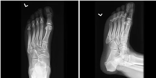
Image 1. Plain radiograph of the left foot. Case courtesy of Frank Gaillard, Radiopaedia.org
What is the diagnosis?
This is a fracture of the base of the fifth metatarsal where the 4th and 5th intertarsal junction occurs, which is classified as a Jones fracture.
Pearl: The location of the fracture in the fifth metatarsal is extremely important and affects treatment. Lawrence and Botte developed classifications which used three zones. Fractures that are more proximal are Zone 1. These are pseudo-Jones or tuberosity avulsion fractures. Zone 2 is the intertarsal junction, These are Jones fractures. Zone 3 is the most distal and commonly involves diaphyseal stress fractures (2).

Image 2: Drawing of the cuboid, fourth and fifth metatarsals. Brown indicates area of pseudo-Jones fracture, red indicates Jones fracture, and blue most commonly involved diaphyseal stress fracture. Drawn by artist and physician Dr. Campbell
What is the mechanism of injury?
Usually either a medially directed force on a plantarflexed foot or a strong sudden adduction/inversion of the forefoot.
Pearl: With a Jones or pseudo-Jones fracture pain should be acute. If a patient’s pain has been chronic this should raise suspicion of a stress fracture (3).
What physical exam findings are expected?
Likely the patient will have difficulty bearing weight and ambulating. There may be swelling and ecchymosis. There is usually tenderness along the base of the fifth metatarsal. Passive inversion and resisted eversion can cause pain.
Which imaging modalities can be used?
X-rays are the way to go. You need a lateral, AP, and oblique images of the foot.
What is the management in the ED?
Control the patient’s pain! Also, with a Jones’ fracture the patient needs to be put in a posterior ankle splint and cannot bear weight for approximately 6-8 weeks. This patient needs to follow up with orthopedics within the next 7 days.
Pearl: While the majority of Jones fractures can be treated nonoperatively, surgical treatment is often recommended to high level athletes to reduce time to heal (4,5).
Pearl: Zone 2 is a vascular watershed area. This can contribute to non-union rates as high as 30% (3).
Pearl: Injuries to Zone 1 can bear weight as tolerated and use a hard sole. Zone 3 fractures are also non-weight bearing and also should be out in a posterior splint.
When do you consult Orthopedics?
Typically Orthopedics consultation is not required for this type of injury. However, Ortho should be consulted whenever there is concern for neurovascular compromise, evidence of an open fracture, or severely displaced fracture.
References
Datir A, Niknejad M, Fahrenhorst-Jones T, et al. Jones fracture. Radiopaedia. Accessed June 1, 2023. https://radiopaedia.org/articles/jones-fracture-1?lang=us
Lawrence SJ, Botte MJ. Jones' fractures and related fractures of the proximal fifth metatarsal. Foot Ankle. 1993;14(6):358-365. https://pubmed.ncbi.nlm.nih.gov/8406253/
Smidt KP, Massey P. 5th Metatarsal Fracture. In: StatPearls. Treasure Island (FL): StatPearls Publishing; May 29, 2023. https://pubmed.ncbi.nlm.nih.gov/31335089/
Cheung CN, Lui TH. Proximal Fifth Metatarsal Fractures: Anatomy, Classification, Treatment and Complications. Arch Trauma Res. 2016;5(4):e33298. https://pubmed.ncbi.nlm.nih.gov/28144601/
Mologne TS, Lundeen JM, Clapper MF, O'Brien TJ. Early screw fixation versus casting in the treatment of acute Jones fractures. Am J Sports Med. 2005;33(7):970-975. https://pubmed.ncbi.nlm.nih.gov/15888715/